Eye scans may spot Parkinson’s years before diagnosis

New technology application designed to help people with Parkinson’s walk more confidently
5th November 2024
Balance Impairment Leading to Falls in Parkinson’s
7th November 2024Eye scans may spot Parkinson’s years before diagnosis

Eye scans may spot Parkinson’s years before diagnosis
Researchers hopeful eye scans will become a pre-screening tool for at-risk people.
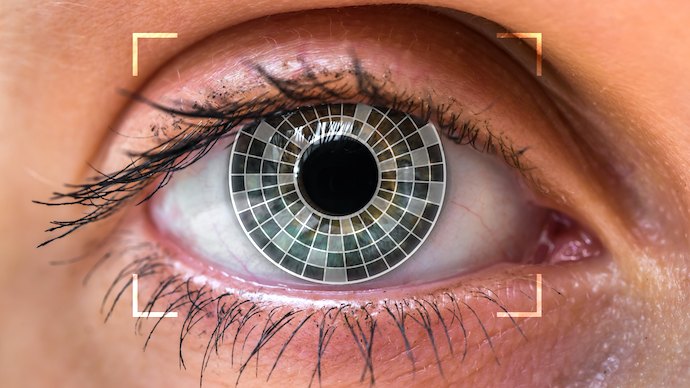
Eye scans may help identify who’s at risk of developing Parkinson’s several years before symptoms first become apparent and doctors can make a diagnosis of the disease, a study suggests.
Researchers observed that two specific structures in the retina – a light-sensitive layer of tissue at the back of the eye – were thinner than normal in people with Parkinson’s, as well as in those who went on to develop the disease up to seven years later.
While it’s still early to know if these measures can be accurate predictors, the findings may be a step forward toward a more rapid diagnosis. For people with Parkinson’s, this could create a better chance of receiving timely intervention.
“I continue to be amazed by what we can discover through eye scans,” said Siegfried Wagner, MD, a researcher at the U.K.’s University College London (UCL) Institute of Ophthalmology and the study’s first author.
“While we are not yet ready to predict whether an individual will develop Parkinson’s, we hope that this method could soon become a pre-screening tool for people at risk of disease,” added Wagner, who also is a clinical research fellow at Moorfields Eye Hospital in London.
The study, Retinal Optical Coherence Tomography Features Associated With Incident and Prevalent Parkinson Disease was published in the journal Neurology.
Diagnosing Parkinson’s can be challenging because its symptoms emerge gradually, sometimes taking years to become noticeable. It would be important to identify signs of the disease early on when intervention would be most effective.
“Finding signs of a number of diseases before symptoms emerge means that, in the future, people could have the time to make lifestyle changes to prevent some conditions arising,” Wagner said.
The researchers already knew that one the retina’s layers, called the ganglion cell–inner plexiform layer (GCIPL), is thinner in people with Parkinson’s than in those without the disease.
Eye scans culled from two databases
Now, they used eye scans available from two large databases, the AlzEye and the UK Biobank, to find other signs that may lead to earlier diagnosis of Parkinson’s. All eye scans were taken using optical coherence tomography (OCT), a technique that offers detailed pictures of the retina.
To make measurements faster and more accurate, the researchers used a software tool for automated segmentation. This refers to a process whereby area boundaries are assigned automatically by a computer program.
Of the 154,830 people with eye scans available from AlzEye, 700 (0.45%) had Parkinson’s. Compared with control individuals who did not have Parkinson’s, those with the disease were older, more likely to be men, and have high blood pressure (hypertension) or diabetes.
After accounting for these factors, the researchers observed that people with Parkinson’s had a GCIPL that was on average 2.12 micrometers (mcm) thinner than that of people without the disease.
Inner nuclear layer also thinner
Another layer known as inner nuclear layer (INL) was also on average 0.99 mcm thinner compared to that of people without Parkinson’s. The researchers noted that the cell bodies of dopaminergic neurons – those that are lost gradually throughout the course of Parkinson’s – sit at the border of the INL.
In the UK Biobank, eye scans were available from 50,405 people, mean age 56.1 years. Of these, 53 (0.1%) went on to develop Parkinson’s at a mean of 7.3 years, after their eye scans.
Both thinner GCIPL and thinner INL were linked to higher chances of developing Parkinson’s in the future.
“Individuals with [Parkinson’s] have reduced thickness of the INL and GCIPL of the retina,” the researchers wrote. “Involvement of these layers several years before clinical presentation highlight a potential role for retinal imaging for at-risk stratification of [Parkinson’s].”
“This work demonstrates the potential for eye data, harnessed by the technology to pick up signs and changes too subtle for humans to see,” said Alastair Denniston, MD, PhD, a consultant ophthalmologist at University Hospitals Birmingham in the U.K. and a study author.
“We can now detect very early signs of Parkinson’s, opening up new possibilities for treatment,” Denniston added.
Sources:
Original article by Margarida Maia, PhD


